Home » Without Label » Upper Leg Tendon Anatomy / Muscles of the Thigh and Gluteal Region - Part 1 - Anatomy ... - Tendons are cords made of tough tissue, and they work as special connector pieces between bone and muscle.
Upper Leg Tendon Anatomy / Muscles of the Thigh and Gluteal Region - Part 1 - Anatomy ... - Tendons are cords made of tough tissue, and they work as special connector pieces between bone and muscle.
Upper Leg Tendon Anatomy / Muscles of the Thigh and Gluteal Region - Part 1 - Anatomy ... - Tendons are cords made of tough tissue, and they work as special connector pieces between bone and muscle.. The vastus laterails works with the other quad muscles to help extend your knee joint. Upper leg tendon anatomy : Rectus femoris these four muscles come together to form a single tendon, which inserts into the patella, or kneecap. It is thin and flattened, broad above, narrow and tapering below. The achilles tendon or heel cord, also known as the calcaneal tendon, is a tendon at the back of the lower leg, and is the thickest in the human.
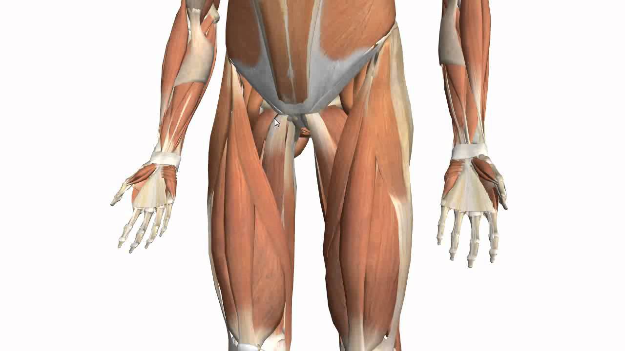
This mri wrist coronal cross sectional anatomy tool is absolutely free to use. The muscles of the thigh and gluteal region are a group of complex muscles that help move and stabilize the lower limb. The thigh is the region between the hip and knee joints. People who play soccer have these specific muscles of the leg very well defined, so they're like a walking anatomy atlas for thigh muscles. The fibers run vertically downward, and end in a rounded tendon, which passes behind the medial condyle.
Muscles of the Thigh Part 2 - Medial Compartment - Anatomy ... from i.ytimg.com The patella is attached to the shinbone (tibia) by the patellar tendon. Upper leg tendon anatomy : Is there an easy way to learn their a. It's the area that runs from the hip to the knee in each leg. Upper leg muscle pain is a very hard pain affect the leg pain as a whole. Your lower leg includes three main muscles, located behind your tibia or shinbone. The achilles tendon or heel cord, also known as the calcaneal tendon, is a tendon at the back of the lower leg, and is the thickest in the human. The achilles tendon or heel cord, also known as the calcaneal tendon, is a tendon at the back of the lower leg, and is the thickest in the human body.
See more ideas about muscle anatomy, leg muscles anatomy, leg muscles.
Upper leg muscle pain is a very hard pain affect the leg pain as a whole. Other muscles of the anterior (front) thigh include the pectineus, sartorius,. Tendons are thick bands of tissue that connect muscles to bone. Upper leg tendon anatomy gelas tuah mei 18, 2021. The thigh has three sets of strong muscles: The vastus laterails works with the other quad muscles to help extend your knee joint. A muscle strain (muscle pull or tear) is a common injury, particularly among people who participate in sports. It serves to attach the plantaris, gastrocnemius (calf) and soleus muscles to the calcaneus (heel) bone. These muscles run from the lower spine. Ebraheim's educational animated video describes muscle anatomy of the thigh. In clinical anatomy the thigh muscles are divided into three groups: Superficial veins of upper limb , anatomy : The iliopsoas muscle flexes your hip, bends your trunk towards your thigh and rotates your thigh bone.
•medial thigh muscles•adductor longus muscle•adductor magnus muscle•adductor. Your upper leg includes seven major muscles. Possibly the most important tendon in terms of mobility is the achilles tendon. Squeeze your knees together and boom, you're contracting the adductors. Possibly the most important tendon in terms of mobility is the achilles tendon.
BIOL 160: Human Anatomy and Physiology | Human anatomy ... from i.pinimg.com Tendons are thick bands of tissue that connect muscles to bone. Related posts of muscle anatomy upper leg. Suspensory ligament of the axilla. The quadriceps tendon attaches the quadriceps muscles to the patella. This is why you have to indicate which biceps you are taking about when discussing one or other of these muscles. Lateral (fibular) collateral ligament (fcl) upper part middle part lower part popliteus tendon (pt) upper part i. Shoulder muscle anatomy image 12 photos of the shoulder muscle anatomy image shoulder muscle anatomy images, shoulder muscle anatomy picture, human muscles, shoulder muscle anatomy images, shoulder muscle anatomy picture. It's the area that runs from the hip to the knee in each leg.
Anatomy the four quadriceps muscles meet just above the kneecap (patella) to form the quadriceps tendon.
The thigh muscles are divided into three compartments: See more ideas about muscle anatomy, leg muscles anatomy, leg muscles. Shoulder muscle anatomy image 12 photos of the shoulder muscle anatomy image shoulder muscle anatomy images, shoulder muscle anatomy picture, human muscles, shoulder muscle anatomy images, shoulder muscle anatomy picture. Upper leg muscle pain is a very hard pain affect the leg pain as a whole. Learn about the muscles, tendons, bones, and ligaments that comprise the knee joint anatomy. It arises by a thin aponeurosis from the anterior margins of the lower half of the symphysis pubis and the upper half of the pubic arch. This important tendon in the back of the calf and ankle stores the elastic. Its muscle belly is on the back aspect of the upper arm. On the medial edge of the posterior thigh is the gracilis muscle. Possibly the most important tendon in terms of mobility is the achilles tendon. It is thin and flattened, broad above, narrow and tapering below. Your upper leg includes seven major muscles. The iliopsoas muscle flexes your hip, bends your trunk towards your thigh and rotates your thigh bone.
It's the area that runs from the hip to the knee in each leg. This mri wrist coronal cross sectional anatomy tool is absolutely free to use. The patella is attached to the shinbone (tibia) by the patellar tendon. Tendons are thick bands of tissue that connect muscles to bone. This important tendon in the back of the calf and ankle stores the elastic.
Sartorius Stock Images, Royalty-Free Images & Vectors ... from thumb7.shutterstock.com A muscle strain (muscle pull or tear) is a common injury, particularly among people who participate in sports. In clinical anatomy the thigh muscles are divided into three groups: They consist of the rectus femoris, vastus intermedius, vastus lateralis and the vastus medialis. This is why you have to indicate which biceps you are taking about when discussing one or other of these muscles. It is thin and flattened, broad above, narrow and tapering below. 430) is the most superficial muscle on the medial side of the thigh. Upper leg anatomy and function the upper leg is often called the thigh. The thigh has three sets of strong muscles:
Squeeze your knees together and boom, you're contracting the adductors.
Other muscles of the anterior (front) thigh include the pectineus, sartorius,. In clinical anatomy the thigh muscles are divided into three groups: It is thin and flattened, broad above, narrow and tapering below. This important tendon in the back of the calf and ankle stores the elastic. Squeeze your knees together and boom, you're contracting the adductors. Lateral (fibular) collateral ligament (fcl) upper part middle part lower part popliteus tendon (pt) upper part i. Concept conceptual 3d illustration fit strong back upper leg human anatomy, anatomical muscle. Notice the upper leg has a biceps muscle just like the upper arm does. Tendons are thick bands of tissue that connect muscles to bone. The patella is attached to the shinbone (tibia) by the patellar tendon. The fibers run vertically downward, and end in a rounded tendon, which passes behind the medial condyle. It serves to attach the plantaris, gastrocnemius (calf) and soleus muscles to the calcaneus (heel) bone. •medial thigh muscles•adductor longus muscle•adductor magnus muscle•adductor.